Urography is an examination used to evaluate the kidneys, ureters and bladder. Excretory urography, also known as intravenous pyelogram (IVP) is performed using conventional x-ray after the intravenous administration of radiographic contrast material. This technique is still performed for pediatric patients and occasionally for young adults.
Computed tomography (CT) urography and magnetic resonance (MR) urography use CT and MR images, respectively, after intravenous contrast material to obtain images of the urinary tract. CT urography (CTU) and MR urography (MRU) are used as primary imaging techniques to evaluate patients with blood in the urine (hematuria), follow patients with prior history of cancers of the urinary collecting system and to identify abnormalities in patients with recurrent urinary tract infections. In addition to imaging the urinary tract, CT and MR urography can provide valuable information about other abdominal and pelvic structures and diseases that may affect them.
MR or CT Urography is done for the following conditions :
Hematuria (blood in urine)
Kidney or Bladder Stones
Cancer of the Urinary Tract
In order to distend your urinary bladder, you may be asked to drink water prior to the examination, and also not to urinate until after the scan is complete. For MR urography, you may have contrast material injected intravenously for the exam, so it is better if you come for the exam in fasting state.
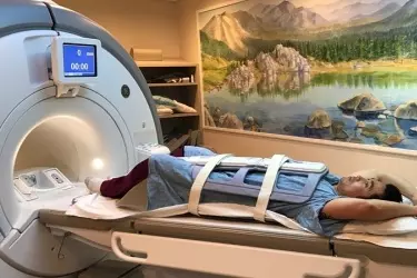
During the MRI exam, you would be in a room which has strong magnetic field. You should not be wearing any jewellery and should inform the technician if you have any kind of implant inside your body. In any case our technician would be checking you with a metal detector before taking you inside the magnet room. The study would take around 15-30 minutes to complete.
You are advised to leave your mobile phone, wallet and all clothes inside a locker which is provided to you. You would be given a hospital gown to wear for the test. There is a humming sound which resembles the sound of a running train while the exam is going on. You would be given ear plugs to minimize this sound. You would be required to go inside a cylinderical tunnel and lie still while the exam is going on. On completion of the test, you can change into your own clothes and leave for your home. The MRI Urography exam will produce several hundred images which will be reviewed by the radiologist and the report would be made available within 24 hours of the scanning.
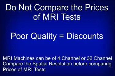
MRI Machines are of two types - Permanent Magnet and Superconducting Magnet. The Permanent Magnet MRI or Open MRI as it is called comes in two strengths - 0.2 Tesla and 0.3 Tesla. The Superconducting Magnet comes in strengths of 1.5 Tesla and 3.0 Tesla. Tesla is the unit for magnetic strength. The common belief is that higher the Tesla, better is the imaging quality but this is not the case. Higher magnetic strength can produce greater noise and result in lower signal to noise ratio. MRI of the Brain, Joints and Spine can be done equally well on a 0.3 Tesla Permanent Magnet as it is done on a 1.5 Tesla Magnet. However inner ear imaging is best done on a 3 Tesla MRI. Many doctors would prescribe MRI scan on a 3 Tesla Machine without knowing the difference in imaging quality. The quality of imaging also depends on the quality of coils, channels in the machine and specialized applications for doing the scanning. Finally the quality of reporting is of paramount importance in any MRI Imaging. At Bharat Diagnostics we have the optimum combination of all these factors. We have all types of machines at different centres. So patients who are claustrophobic, are advised to get their MRI on open magnet of 0.3 Tesla. Patients who require extensive testing like MRI brain and whole spine would do better to go for 3 Tesla as the imaging time is lesser.
The cost of MRI Spine for each level - Cervical, Dorsal or Lumbar at our centres is in the range of Rs. 7000 - 7500. If contrast is needed then additional amount of Rs. 3500 is charged for the test. MRI Screening for each level would cost Rs. 2500 - 3500. MRI Screening for the whole spine can be done in the range of Rs. 7500 - 8500.
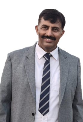
Dr Rajesh Choudhary has more than 20 years of experience in the field of Radiology. Dr Choudhary has been trained in the latest radiological techniques from Sardar Patel Medical College, Bikaner and PGI Chandigarh.

Dr Shruti Sangwan has been trained at Lady Hardinge Medical College, Maulana Azad Medical College, PGI Chandigarh and Artemis Hospital Bhiwadi in the field of Radiology and has more than 18 years of experience.
